Learning objectives
Brainstem (Rhomboencephalon)
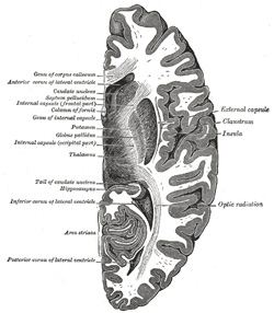
|
|
Brainstem Function
The Brainstem carries all descending and ascending fibres from the brain to the spinal cord or cerebellum. This includes motor fibres down from motor cortex to spinal cord in the corticospinal tract. These are carried on the front "ventral surface" in the cerebral peduncles at midbrain level, the pyramids in the pons and medulla. The main vascular supply to the brainstem also lies anteriorly on the ventral surface with the vertebral arteries, basilar artery and the posterior cerebral artery. Branches of these provide branches which supply the brainstem. The brainstem also contains the reticular activating system associated with arousal is found throughout. As its name suggests is a network of interconnecting cells with different functions. Circulation, respiration, sleep and wakefulness.
Cranial nerves and their nuclei
- Midbrain III and below this IV
- Pons V,VI,VII,IX
- Medulla IX, X, XI,XII
Other contents of brainstem
- Sympathetic outflow
- Afferent from and efferents to cerebellum
- Corticospinal fibres
- Corticobulbar fibres
- Ascending sensory - dorsal and spinothalamic
- Reticular activating system
- Olivocerebellar fibres from inferior olive to cerebellum
- Rubrospinal from red nucleus
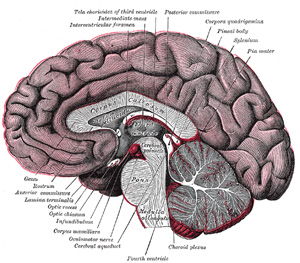
Midbrain (Mesencephalon)
Midbrain lies below the diencephalon. Ventrally (in front) are two large cerebral peduncles containing the fibres of the internal capsule on either side of the midline in which lie axons on their way from the cerebral cortex to the brainstem or spinal cord including the pyramidal tracts. The emerging oculomotor nerves emerge from between these. On the dorsum lies 4 small hillocks of the two super colliculus (reflex eye movements) above and the two inferior colliculi (auditory pathway) below. The trochlear nerve emerges just below the inferior colliculus and is the only cranial nerve to emerge posteriorly. It contains the Cranial nerve nuclei 3 and 4. Smallest part of the brainstem and is continuous with the diencephalon above and the pons below. Contains the cerebral aqueduct between the tectum and tegmentum. Rather than medial and lateral contents the anatomy is split into those structures at the level of the superior and then inferior colliculus. The Midbrain contains
Midbrain at Super colliculus level
- Superior colliculus lies dorsally as a small hillock
- Cerebral aqueduct surrounded by periaqueductal grey matter
- Oculomotor nucleus on either side of midline in front of aqueduct. Supplies ipsilateral extraocular muscles and pupil. Fibres run forwards and exit anteriorly between cerebral peduncles
- Red nucleus lies on either side
- Substantia nigra lies between main brainstem and cerebral peduncles. Pigmented. Important in Parkinson's disease.
- Cerebral peduncles lie anteriorly and contain the corticonuclear fibres medially and the corticospinal fibes laterally
- Medial longtitudinal fasciculus
- Medial lemniscus
- Dentatothalamic tract
- Medial Geniculate body lies laterally
- Reticular activating system
Midbrain at Inferior colliculus level
- Inferior colliculus lies dorsally
- IVth nerve nucleus is found at this level just on either side of midline
Vascular supply is from the basilar artery and the posterior cerebral artery. Infarction can be medial, laterally, rostral or caudal and can extend to thalamus and cortical areas supplied by PCA. In the midline posteriorly lies the aqueduct carrying CSF from IIIrd to IVth ventricle. Surrounded by grey matter (cell bodies) called the periaqueductal grey matter which is involved in pain perception and stress responses. Midbrain also colourful as more anteriorly it has the red nucleus and the substantia nigra.
Pons
Ventrally rather bulbous due to large cell groups and fibres running transversely between large pontine nuclei and the cerebellum. Laterally lies the bulging middle cerebellar peduncle. The pontine nuclei mediate fibres coming from cerebral cortex to the cerebellum. The VI, VII, VIII and V nerves all leave ventrally. As its name suggests it is bridge like and like any bridge it is a major crossover centre for fibres carrying signals bidirectionally from above and below and communication with the cerebellum. Like any bridge it covers the CSF filled 4th ventricle which separates it from the cerebellum with its vermis and hemispheres which sit tightly behind.
The Pons contains the Corticospinal fibres, Corticopontine fibres, Pontocerebellar fibres and Raphe nuclei. The lower Pons contains the Nucleus of VI, Nuclei of VII, Facial nerve, Superior salivatory Gustatory. The facial nerve and abducent nerve are closely associated as the exiting VII nerve loops around the nucleus of VI. Also contained is the Medial lemniscus (carries contralateral dorsal column), Spinothalamic tract (carries pain and temperature) and Middle cerebellar peduncle which contains fibres from C/L pons to cerebellum.
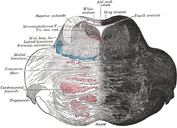 |
Medial structures
- Medial longitudinal fasciculus
- VI (Abducent) nerve nucleus and fibres
- Genu of VII (Facial) closely associated with VI
- Medial lemniscus (carries contralateral dorsal column)
- Corticospinal tract
Lateral structures
- Facial nucleus VII and nerve fibres
- Spinal nucleus and tracts of V
- Lateral spinothalamic tract (spinal lemniscus)
Vascular supply
From the basilar artery which lies anteriorly and provides median and paramedian perforators. Short circumferential branches irrigate more laterally and laterally including the middle cerebral peduncle is supplied by branches of the superior cerebellar and anterior inferior cerebellar arteries.
Medulla
The medulla contains a host of nuclei all tied up with fairly primitive functions such as cardiorespiratory, vomiting centres etc. It also contains the reticular activating system. Medially to the olive and near the midline is the medial lemniscus that carries dorsal column sensation to the thalamus. Vascular supply is by branches of the vertebral artery including the posterior inferior cerebellar artery. PICA as we know classically irrigates lateral medulla. Anteriorly there are median and paramedian perforators from the vertebrals. Lateral medullary syndrome is more commonly due to vertebral occlusion than PICA occlusion of itself.
Medial structures
- Hypoglossal nerve nucleus controls tongue muscles and the nucleus lies near the midline and exists the posterior fossa through the hypoglossal formina. Each hypoglossal receives bilateral projections from the motor cortex. Unilateral weakness causes the tongue to bend to the weak side. Tongue can be weak and also can be apraxic.
- Medial lemniscus with crossed fibres from the dorsal column (gracile and cuneate nuclei) on the way to the ipsilateral thalamus
- Pyramids lie on either side of the midline on the ventral surface and contain the corticospinal motor tracts which are decussating.
Lateral structures
- Nucleus ambiguus : Receives bilateral cortical innervation from the primary motor cortex. Efferents to IX and X and XI. Concerned with swallowing. Descending nucleus of V: which has its cell bodies within the trigeminal ganglion. Dorsal motor nucleus of X : preganglionic parasympathetic to nerves IX and X. Supplies heart, lungs and abdominal organs.
- Nucleus solitarus : Lies in the medulla and receives input from VII, IX and X. Part of it is called the gustatory nucleus concerned with taste. Fibres pass from here to the venteroposteromedial nucleus of thalamus.
- Laterally to the pyramids is the bulge of the inferior olivary nucleus which sends fibres to the cerebellum
- Vestibular nuclei : four of these lie in floor of the IVth ventricle. Cochlear nuclei : closely associated with vestibular nuclei
- Inferior cerebellar peduncle with dorsal spinocerebellar, olivocerebellar and cuneocerebellar tracts
- Lateral spinothalamic tract (spinal lemniscus)
- Spinal nucleus of V: Fibres concerned with touch, pain and temperature from the face goes to the spinal nucleus of V. Descend from pons down to the medulla.
Vascular supply is from the posterior inferior cerebellar artery which supplies the lateral medulla as well as the underside of the cerebellum. Occlusion to the PICA or the vertebral itself will cause an ipsilateral Lateral medullary syndrome. Medially the anterior spinal artery if occluded can cause a medial medullary syndrome. Penetrating branches of the vertebral can also supply structures.
| Note: The plan is to keep the website free through donations and advertisers that do not present any conflicts of interest. I am keen to advertise courses and conferences. If you have found the site useful or have any constructive comments please write to me at drokane (at) gmail.com. I keep a list of patrons to whom I am indebted who have contributed. If you would like to advertise a course or conference then please contact me directly for costs and to discuss a sponsored link from this site. |