Learning objectives
|
Introduction
Clinical assessment and examination commences with the first responder or layperson to quickly identify suspected stroke. The history is key but this is then followed by a rapid and focused clinical exam. For those not trained there are several easy to perform screening tests. There are three levels of identification that need to be spot on to acts as sieves to identify and find all of those who may have a stroke. It is important to note that no test or assessment is 100% and there must always be a way to enable discussion between first responder or emergency department and specialist when needed.
| Level | Tests and Assessment |
|---|---|
| Prehospital | In the UK the FAST test is the key test for both laypersons and First responders. |
| Emergency Department | In the ED the ROSIER scale may be used. This may be bypassed where there is direct first responder to stroke team communications |
| Stroke Team | Expert focused assessment using NIHSS and clinical acumen. Initial contact may be with Specialist nurse and rapid escalation. |
How it goes wrong
We still see that every once in a while, patients have a delayed referral. There is some cognitive dissonance issues. Possible reasons
| Issue | Comments |
|---|---|
| They are too young to have a stroke | All age groups can have a stroke. We do see teenagers and young adults with big strokes. If the person has a typical stroke syndrome then you must escalate. In a stroke like clinical syndrome do not diagnose Migraine or Seizure or non-stroke pathology without senior discussion with stroke physician |
| The CT is normal | The CT is often normal in an Ischaemic stroke under 6 hours old. Signs on CT are subtle at this time. |
| The patient tells me he is fine or is getting better | Unless the patient is now up and walking about with no deficit then assume they are not back to normal. Patients with Right cortical strokes often tell people they are fine. It’s part of their anosognosia. |
Prehospital Stroke Assessment (Bystander/First responder)
Early recognition is key to early treatment and therefore there has to be a good level of bystander ability to recognise a stroke and act quickly. There has been a huge amount of publicity to make the public and other healthcare workers more aware of the early signs of possible stroke. Various assessment tools have been suggested for public and paramedical staff. For the sake of interest I have included a few of them here but the one which is used nationally is the FAST test which we will go on to to discuss.
Prehospital use: The Cincinnati Prehospital Stroke Scale
Tests three things - face, arm droop and speech and as you will see it is very similar to FAST. It was based on a simplification of the National Institutes of Health (NIH) Stroke Scale.
| Cincinnati Prehospital Stroke Scale | |
|---|---|
| Facial droop | Have the person smile or show his or her teeth. If one side doesn't move as well as the other so it seems to droop, that could be sign of a stroke. Normal: Both sides of face move equally or Abnormal: One side of face does not move as well as the other (or at all) |
| Arm drift | Have the person close his or her eyes and hold his or her arms straight out in front for about 10 seconds. If one arm does not move, or one arm winds up drifting down more than the other, that could be a sign of a stroke. Normal: Both arms move equally or not at all. Abnormal: One arm does not move, or one arm drifts down compared with the other side |
| Speech | Have the person say, "You can't teach an old dog new tricks," or some other simple, familiar saying. If the person slurs the words, gets some words wrong, or is unable to speak, that could be sign of stroke. Normal: Patient uses correct words with no slurring. Abnormal: Slurred or inappropriate words or mute |
| Assessment | The CPSS was found to have excellent reproducibility among prehospital personnel and physicians. It has good validity in identifying patients with stroke who are candidates for thrombolytic therapy, especially those with anterior circulation stroke [Kothari et al. 1999]. |
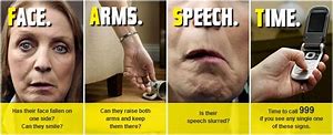
Prehospital use: Face Arm Speech Test (FAST)[1]
This is very similar to CPSS. Suspect stroke if any of the following is answered “yes”:
| Face Arm Speech Test (FAST) | |
|---|---|
| Face | Facial movements - Ask patient to smile or show teeth. Look for new asymmetry. Is there an unequal smile or grimace or obvious facial asymmetry. |
| Arm | Arm movements : Lift the patient's arms together to 90 degrees if sitting, or 45 degrees if supine. Ask him to hold that position for 5 sec and then let go. Does one arm drift down or fall more rapidly? |
| Speech | Speech impairment : Look for new disturbances in speech. Look for slurred speech and word finding difficulties. |
| Test | test all three and ring 999/911 if any one of these is positive |
Paramedics using the Face Arm Speech Test [1] achieved high levels of detection and diagnostic accuracy of stroke
List of Prehospital Acute Stroke Screening Tools
- Cincinnati Prehospital Stroke Scale (CPSS)
- Face Arm Speech Test (FAST)
- Los Angeles Prehospital Stroke Screen (LAPSS)
- Melbourne Ambulance Stroke Screen (MASS)
- National Institutes of Health Stroke Scale (NIHSS)
- Recognition of Stroke in the Emergency Room (ROSIER)
Emergency Dept Stroke Screening: ROSIER Scale [2]
Once patients get to the ED where they can be seen by a physician then the ROSIER scale (Recognition of Stroke in the Emergency Room) has been advocated and is widely used. The ROSIER scale as detailed is simply an enhanced FAST test that subtracts from the stroke score if there is documented hypoglycaemia or a seizure at symptom onset. It also includes visual loss and leg weakness within the clinical criteria. As a screening test more recent studies have shown that it does not add much beyond the sensitivity and specificity of the simpler FAST test (Whitely et al. 2011) and it may just delay escalation to the stroke service. Locally we would alert stroke team on basis of FAST and initial clinical assessment. ROSIER is included here for your information and to be aware of it as it is on the stroke proforma of many hospitals. It may well be used in your local hospital. There are other screening tools.
| ROSIER SCALE | |
|---|---|
| Has there been loss of consciousness or syncope? | Y(-1) N (0) |
| Has there been seizure activity? | Y(-1) N (0) |
| Is there a NEW ACUTE onset (or on awakening from sleep)? | Y(-1) N (0) |
| I. Asymmetric facial weakness | Y(+1) N(0) |
| II. Asymmetric arm weakness | Y(+1) N(0) |
| III. Asymmetric leg weakness | Y(+1) N(0) |
| IV. Speech disturbance | Y(+1) N(0) |
| V. Visual field defect | Y(+1) N(0) |
| Total score = (-2 to 5) | |
| If total score > 0 (1 to 6) a diagnosis of acute stroke is likely. If total scores 0, -1 or -2 stroke unlikely but is not excluded and patient should be discussed with the stroke team. | |
If at this stage an acute stroke with onset within past 4-4.5 hours is diagnosed please alert stroke team urgently and arrange an urgent head CT.
Screening tests
|
| ROSIER (90% CI) | CPSS (95% CI) | FAST (95% CI) | LAPSS (95% CI) |
|---|---|---|---|---|
| Sensitivity | 93 (89-97) | 85 (80-90) | 82 (76-78) | 59 (52-56) |
| Specificity | 83 (77-89) | 79 (73-85) | 83 (77-89) | 85 (80-90) |
| Positive Predictive value | 90 (85-95) | 88 (83-93) | 89 (84-94) | 87 (82-92) |
| Negative Predictive Value | 88 (83-93) | 75 (68-82) | 73 (66-80) | 55 (48-62) |
Stroke Team : Clinical Examination
Recognising typical pattern combinations aids diagnosis and is key to a rapid and accurate diagnosis. Strokes are vascular syndromes and non haemorrhagic strokes must match the syndrome of the 'culprit' artery occluded. The examination below is the exam to be done when there is no time pressure for urgent therapies. Otherwise the general examination is the NIHSS scale.
| Clinical Signs |
|---|
|
Clinical examination in stroke disease must define the deficit and resultant disabilities and also help focus investigations on the underlying aetiology. Must constantly ask "Where and why". In the acute situation when time is short the NIH is a useful way to do a methodical neurological examination that covers patterns of deficits seen in stroke. One must be doing this routinely. It is detailed in the appendix at the back.
General Examination in Stroke
The general examination of a stroke patient is not confined to one system but should be from top to toe covering the neurological deficit and any suspected causative features or stroke complications.
Coma
Coma in stroke disease suggests a major bleed or infarct with raised intracranial pressure perhaps due to hydrocephalus or cerebral oedema. Most stroke patients are otherwise alert even with marked neurology. In a younger patient under 60 with a malignant MCA infarct progressive coma may indicate urgent hemicraniectomy or developing hydrocephalus. In a patient with a cerebellar bleed urgent clot evacuation is needed or ventricular drainage for obstructive hydrocephalus. Somnolescence is different to coma - the patient appears to be sleeping rather than coma e.g. moves in bed, gets position of comfort but unrousable. Consider bilateral thalamic or other high brainstem infarction.
Causes of coma to be considered in a stroke patient
- Stroke with raised ICP due to cerebral oedema, haematoma or hydrocephalus
- Post Seizure or Non convulsive status
- Hypoglycaemia - always test if a possibility
- Bilateral brainstem/thalamic strokes
- Sedatives taken e.g. Benzodiazepines, codeine, opiate skin patches
- Stroke + sepsis/metabolic cause e.g. Severe hyponatraemia
Always identify the cause an decide whether action is needed or not.
Appearance
- Habitus: Marfan's disease, Ehlers-Danlos syndrome
- Pregnancy is also a risk factor
- Acromegalic or Cushingoid features may suggest endocrine hypertension
- Nicotine staining (in heavy smokers always think is this carotid disease)
Hands
- Splinter haemorrhages may suggest endocarditis or a manual worker (commoner in dominant hand),
- Clubbing may suggest cyanotic congenital heart disease or endocarditis. Might suggest cerebral metastases from a lung tumour masquerading as a stroke.
- Palmar erythema, spider naevi
- Dupuytrens may suggest alcoholism with risks of ischaemic and haemorrhagic stroke
- Arachnodactyly "spider fingers" seen in Marfan's and some connective tissue diseases with a higher risk of aortic incompetence, mitral valve prolapse and arterial dissection.
Face
Facial Pulses
- Look for temporal artery pulses and tenderness which may suggest GCA
- Dynamic testing of facial pulses may help suggest if ipsilateral carotid occluded. This will cause external carotids to retrogradely supply INC territory. One place where the two systems meet is at the eye where the ophthalmic artery supplies. as mentioned above in the discussion about internal carotid disease. An absent or tender temporal artery is one of the criteria in the diagnosis of giant cell arteritis. Always document where there is a new headache or visual loss even transient resembling amaurosis fugax. Seen in those over 50 and can rarely cause stroke. Check an ESR urgently and consider steroids and ophthalmic referral for confirming diagnosis and temporal artery biopsy.
Eye
- Pallor from anaemia
- Yellow discolouration from jaundice
- Xanthelasma suggesting dyslipidaemia
- Small miosed pupil and mild ptosis may suggest a Horner's syndrome due either to carotid artery dissection or an apical lung tumour or a lateral medullary infarct.
- Complete or almost complete ptosis and dilated pupil pointing down and out suggests an oculomotor palsy in the midbrain or along its path. In some cases e.g. diabetes the pupil will remain intact.
Ptosis - can be due to weakness of 3 muscles
- Levator palpaebrae supplied by III e.g. midbrain infarction
- Muller's muscle supplied by sympathetic e.g. Horner's syndrome e.g. Lateral medullary infarction
- Frontalis supplied by VII and is least important and is not affected in UMN VII and so unaffected in stroke disease,
- Causes of Ptosis: IIIrd nerve (Complete), Horner's syndrome (Partial), Myasthenia gravis, Botulism, Myopathies, soft tissue problems
Visual fields
There are many ways taught to assess visual fields. The confrontation method with opposing eyes closed and fingers moving in from quadrants is absolutely fine. Mostly we are confirming a homonymous hemianopia and one way is to test each eye in turn and show from 1 to 4 fingers in each eye quadrant in turn and see if the patient can appreciate them. If there is a field defect assess for macular sparing by assessing central vision. If there is no field defect then look for visual extinction by presenting simultaneous wiggling fingers in the laterally in each patient and seeing if both are appreciated. Assess visual acuity using Snellen chart if possible. Alternatively using available printed material and stating the best that a patient can do is often sufficient. e.g. can read newspaper small print at 30 cms in both eyes
Cardiorespiratory Examination
Pulse
- Irregularly irregular pulse to look for AF. If suspected get an ECG to confirm.
- Plateau pulse of Aortic stenosis
- Collapsing pulse of AR
- Jerky pulse of hypertrophic cardiomyopathy may also be useful.
Blood pressure
An uncontrolled BP > 185/110 mmHg is a contraindication to thrombolysis. Blood pressure is a major risk factor for both Ischaemic and haemorrhagic stroke. Blood pressure variability may also be important. Both systolic and diastolic values are important though diastolic value tends to fall with age.
Jugular venous pressure
- Elevated JVP can suggest congestive cardiac failure or volume overload.
- SVC obstruction
Chest
- Midline sternotomy - valve replacement or repair or coronary artery bypass grafting or cardiac transplant.
- Pacemaker or AICD scar left or less commonly right infraclavicular fossa.
Palpation of the chest.
- Palpable thrills
- Left sternal heave representing RV hypertrophy and Pulmonary hypertension
- Dyskinetic rocking motion - LV aneurysm
Apex beat
- Any displacement which may suggest cardiac enlargement.
- A palpable tapping S1 may be felt at the apex or medial to it suggesting mitral stenosis
- A heaving apex might suggest aortic stenosis
- Dynamic volume overloaded ventricle may suggest aortic incompetence
Auscultation of the heart
- Systolic Murmur
- Diastolic murmur
- Pericardial sounds
- Metallic valves
Carotid artery
Some stroke books say not to bother as stenosis may be silent and bruits may not mean stenosis and the decision to scan for disease should be based on symptomatology. Still it's hard not to. If there is suspected occlusion of the internal carotid then external carotid flow may increase on the affected side and temporal and facial pulses may increase unilaterally as external carotid flow increases. A bruit over the carotid artery, eyeball, and the skull may actually suggest contralateral carotid occlusion due to increased flow on the side without the occlusion (Fisher's sign). To determine an internal from an external bruit, transiently compress the facial and temporal arteries this increases an internal carotid bruit and decreases an external one. Not part of the examination but as an aside avoid carotid sinus massage in any patient likely too have stroke disease. I would avoid it in any patient over 50. Concerns about dislodging plaques. A normal carotid doppler may make it a less risky procedure. There may even be a large scar running along the edge of the sternomastoid suggesting previous carotid endarterectomy.
Peripheral vascular disease
If the feet are cold and legs hairless then wise to assess peripheral pulses which may suggest peripheral vascular disease. Check ankle-brachial pressures. The blood pressure is reduced at the ankles if there is proximal significant narrowing.
Respiratory examination
- Lung bases for signs of pulmonary oedema
- Signs of long consolidation suggesting pneumonia
- Pleural effusion
- Consolidation
- or stony dullness and reduced breath sounds suggesting pleural effusion with its many causes.
Abdominal and Genital examination
- In the young patient angiokeratomas around the umbilicus might suggest Fabry disease.
- Audible renal bruit may suggests renal artery stenosis and a cause of hypertension
- Enlarged kidneys in adult polycystic kidney disease causing chronic hypertension and well known association with Berry aneurysm and SAH
- Young patient to look for radiofemoral delay due to Coarctation of the aorta. In the past syphilis was the scourge or neurologists and could cause basically any stroke symptom. Indeed Brocas's dysphasic patient had dysphasia due to a gumma and not a stroke. At the stage of neurological disease there is usually no genital evidence of primary or secondary syphilis. Ulcers on the genitals however may be due to Behcet's disease. HIV should always be considered in all sexually active patients.
Cortical dysfunction
| Lobe | Clinical |
|---|---|
| Prefrontal | Disinhibition, poor judgment, Emotional lability |
| Frontal |
|
| Parietal |
Bilateral Parietal Function
Non-Dominant Parietal
Dominant Parietal
|
| Temporal |
|
| Occipital |
|
Assessing Motor Function
Inspection
Take care to look for wasting. Look at quadriceps and palpate muscle bulk. Specifically look at the thenar and hypothenar eminence, 1st dorsal interosseous, deltoids, extensor digitorum brevis. On the face temporal wasting. Additionally look for scars, pressure, vascular and neuropathic ulcers. Always lift the legs and inspect the underside to see heels and between toes for lesions.
Tone
Assess upper limb tone in the wrist and elbow. Get the patient to relax and distract with irregular movements. Different types of altered tone are associated with disease., In the acute stroke the limbs are flaccid and hyporeflexic but this changes with time with increasing tone and extensor plantars and increased reflexes
Spasticity
increased tone seen with pyramidal weakness classically reduces as the joint is moved. High tone often gives way like a penknife and has been called "clasp knife". Best seen in forearm supination in upper limb and knee flexion in lower limb., Extrapyramidal: Lead pipe rigidity as tone is constant through range of movement. Becomes cogwheel when there is overlying tremor. This is seen with Parkinson's disease. Gegenhaltan: an erratic increase in tone in those with frontal lobe dysfunction also called "paratonia"
Power
Weakness in stroke disease follows a particular pattern. Weakness is predominantly in the upper limb extensors and the lower limb flexors. This pyramidal weakness disparity gives the particular flexed upper limb and extended lower limb appearance seen in stroke patients. Without strength in lower limb extensors to hold the knee straight walking would be much more difficult.
Grading power - can be again altered into + or -
- 1. No contraction
- 2. Flicker of contraction
- 3. Active movement no gravity
- 4. Active movement against gravity
- 5. Active movement against gravity and resistance
- 6. Normal power
Reflexes described as 0, +, ++, +++, ++++
Coordination
Finger nose test Ask the patient to touch nose and then your finger making sure the patient extends the full extension of elbow and back. Do this on both sides. There may be evidence of past pointing due to misjudgment of distance and speed. It may suggest cerebellar disease. Heel-shin test: Patient runs heel down the opposite shin and lifts off back onto the patella. Notes speed and coordination.
Heel toe walking: An old sobriety test as alcohol affected cerebellar function. Useful in subtle cases. It does assume the patient has a minimal pyramidal weakness and used really in mobile patients with subtle signs. Romberg's test Used to assess for a sensory ataxia. Usually seen in dorsal column disease where the patient has poor proprioception and closing the eyes leads to falling and loss of position sense
Assessing Sensory Function
Much of this has been covered under parietal lobe function. Sensory loss tends to be complete along one side with large total anterior circulation strokes, thalamic strokes or those that take out the thalamocortical fibres. Look for Extinction when stimuli are presented bilaterally. See Parietal lobe higher functions for more information
Oxford Community Stroke Project classification
This is a useful and commonly used bedside classification for stroke. See Bamford/OCSP Classifications.
Next: >> Using the NIHSS |
References
- [1] Harbinson J et al. Diagnostic Accuracy of Stroke Referrals From Primary Care, Emergency Room Physicians, and Ambulance Staff Using the Face Arm Speech Test. Stroke. 2003;34:71-76
- [2] Nor AM et al. The Recognition of Stroke in the Emergency Room (ROSIER) scale: development and validation of a stroke recognition instrument. Lancet Neurol. 2005 Nov;4(11):727-34.
| Note: The plan is to keep the website free through donations and advertisers that do not present any conflicts of interest. I am keen to advertise courses and conferences. If you have found the site useful or have any constructive comments please write to me at drokane (at) gmail.com. I keep a list of patrons to whom I am indebted who have contributed. If you would like to advertise a course or conference then please contact me directly for costs and to discuss a sponsored link from this site. |