Learning objectives
- Role of the midbrain and its anatomy
- Stroke types and appearance
- Management
Midbrain (Mesencephalon)
- Midbrain lies below the diencephalon. Ventrally (in front) are two large cerebral peduncles containing the fibres of the internal capsule on either side of the midline in which lie axons on their way from the cerebral cortex to the brainstem or spinal cord including the pyramidal tracts. The emerging oculomotor nerves emerge from between these.
- On the dorsum lies 4 small hillocks of the two super colliculus (reflex eye movements) above and the two inferior colliculi (auditory pathway) below.
- The trochlear nerve emerges just below the inferior colliculus and is the only cranial nerve to emerge posteriorly. It contains the Cranial nerve nuclei 3 and 4. Smallest part of the brainstem and is continuous with the diencephalon above and the pons below.
- Midbrain contains the cerebral aqueduct between the tectum and tegmentum. Rather than medial and lateral contents the anatomy is split into those structures at the level of the superior and then inferior colliculus. The Midbrain contains
Schematic of MidBrain anatomy
 |  |
| Superior Colliculus | Inferior Colliculus |
|---|
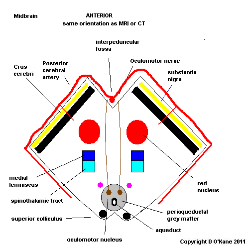
Midbrain at Super colliculus level
- Superior colliculus lies dorsally as a small hillock
- Cerebral aqueduct surrounded by periaqueductal grey matter
- Oculomotor nucleus on either side of midline in front of aqueduct. Supplies ipsilateral extraocular muscles and pupil. Fibres run forwards and exit anteriorly between cerebral peduncles
- Red nucleus lies on either side
- Substantia nigra lies between main brainstem and cerebral peduncles. Pigmented. Important in Parkinson's disease.
- Cerebral peduncles lie anteriorly and contain the corticonuclear fibres medially and the corticospinal fibres laterally
- Medial longtitudinal fasciculus
- Medial lemniscus
- Dentatothalamic tract
- Medial Geniculate body lies laterally
- Reticular activating system
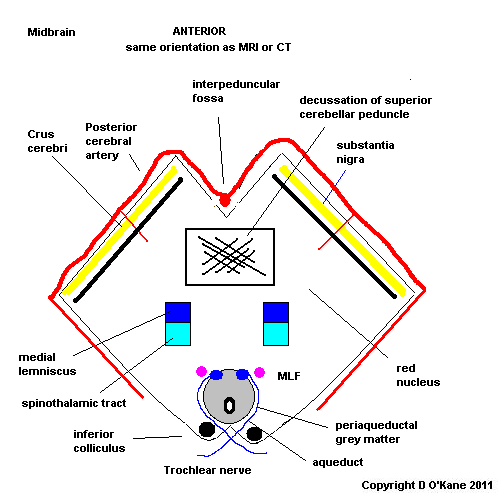
Midbrain at Inferior colliculus level
- Inferior colliculus lies dorsally
- IVth nerve nucleus is found at this level just on either side of midline
In the midline posteriorly lies the aqueduct carrying CSF from IIIrd to IVth ventricle. Surrounded by grey matter (cell bodies) called the periaqueductal grey matter which is involved in pain perception and stress responses. Midbrain also colourful as more anteriorly it has the red nucleus and the substantia nigra.
Vascular supply
- Vascular supply is from the basilar artery and the posterior cerebral artery. Infarction can be medial, laterally, rostral or caudal and can extend to thalamus and cortical areas supplied by PCA.
 |  |
Brainstem Strokes
- The brainstem is vulnerable to both ischaemic and haemorrhagic strokes. Outcome is very variable depending if strokes are unilateral or bilateral. What is important to note is that despite the narrow brainstem the arterial supply is usually from the vertebral or basilar arteries and lesions tend to respect and not cross the midline.
- It is however possible for a very diseased basilar and especially a top of the basilar syndrome to cause multiple bilateral and asymmetrical strokes. Brainstem stroke is not a term to be used only in its most general sense. Cranial nerves and other signs should help one to localise to midbrain, pons and medulla and MRI will support this and the specificity of the location - ventral or dorsal pons etc. Upper or lower midbrain, medial or lateral medulla.
- In the 19th century when stroke location and clinical appearance could only be precisely diagnosed by post mortem French neuroanatomists described and named a collection of brainstem strokes and their clinical signs. These are often used in texts. However, the eponymous terms are not important if the understand how the anatomy and clinical correlate.
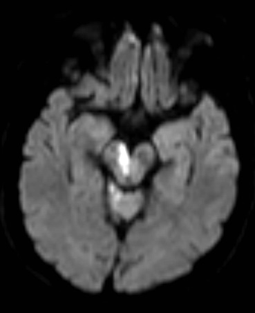
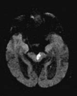
Midbrain Infarction
Right medial midbrain infarct - patient had a right complete IIIrd nerve palsy and left sided hemiparesis. Also known as Weber's syndrome
| Note: The plan is to keep the website free through donations and advertisers that do not present any conflicts of interest. I am keen to advertise courses and conferences. If you have found the site useful or have any constructive comments please write to me at drokane (at) gmail.com. I keep a list of patrons to whom I am indebted who have contributed. If you would like to advertise a course or conference then please contact me directly for costs and to discuss a sponsored link from this site. |