Learning objectives
|
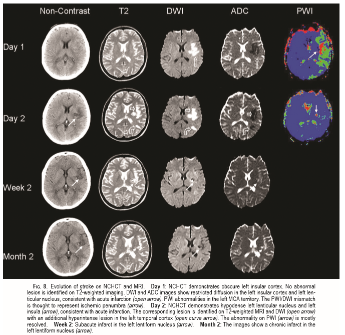
Introduction
Both magnetic resonance imaging (MRI) and computed tomography (CT) have contributed to enormous leaps in neuro-imaging over the past few years. The importance of imaging can be underlined by the 2010 paper which looked at question "Does this patient have a haemorrhagic stroke?: clinical findings distinguishing haemorrhagic stroke from ischaemic stroke." The authors looked at 19 prospective studies meeting inclusion criteria were identified (N = 6438). The conclusion was that in patients with acute stroke, certain findings accurately increase or decrease the probability of intracranial haemorrhage, but no finding or combination of findings is definitively diagnostic in all patients, and diagnostic certainty requires neuroimaging [1]. Rapid multimodal imaging is essential in the workup and management of acute ischaemic stroke. Early parenchymal findings on non-contrast computed tomography or standard magnetic resonance imaging are used to triage patients for intravenous thrombolysis and thrombectomy to provide insight on prognosis. CT without contrast is generally the commonest modality used in the acute stroke setting. Some units use CT with angiography in all patients or where large vessel obstruction or dissection may be considered. Other units outside the UK have adopted MRI as their standard imaging modality. Initially, MRI was extremely motion-sensitive and associated with long acquisition times, so it was difficult to perform in a clinical setting of stroke. The development of faster imaging techniques such as echoplanar imaging rendered it possible to perform an appropriate stroke MR protocol in 20 min or less. MRI is sensitive and relatively specific in detecting changes that occur after such strokes. Higher strength magnets (1.5-3.0 T field strength) give better images. The limitation is the list of contraindications and the fact that those needing monitoring can be difficult along with high cost, longer scanning duration, and decreased sensitivity of subarachnoid haemorrhages.
Working with Neuroradiology
The key people involved are the radiologist and the radiographer. The concern of the radiologist and radiology department is being overwhelmed with unneccesary imaging that takes up scanning time and needs detailed and timely reporting. They often feel obligated to act as gate keepers to scanning. The radiographer is often one of the unsung heroes of stroke care. Radiographers are a vital part of the specialist stroke care team. Patients with acute brain attack require rapid access to high quality and appropriate imaging in order to diagnose the type of stroke and to allow for early intervention, such as thrombolysis and thrombectomy. It is important to build trust between radiology departments and acute stroke services and this can provide efficiencies for both services. If neuroradiologists trust the stroke physicians to request scans through working with radiographers in a way that minimises waste of resources and provides optimal diagnostic work up then this can help to avoid interrupting radiologists to get tests done in the majority of patients. Radiographers undertaking this work not only have highly specialist skills in imaging modalities, but are experienced in the care and techniques required for scanning acutely ill patients who require urgent assessment during a critical period of the care pathway. They may also be able to undertake the further imaging techniques such as CT Angiography and CT perfusion imaging. There will also be a need for MR scanning, specifically for TIA including Diffusion Weighted Imaging (DWI), MR perfusion, and, in addition, Magnetic Resonance Angiography (MRA), Contrast Enhanced MRA (CEMRA) if appropriate, and Carotid USS Imaging[2].
Both CT Brain Imaging and MR Brain Imaging have greatly helped to focus upon and improve clinical care of patients who suffer strokes. Even still with these wonderful modalities they often raise new questions and dilemmas and uncertainties. It is important to always treat the patient and not the scan. Strokes can mimic tumours and vice versa. MRI technology provides us with a huge amount of information some of which we do not know the significance of. We need to use such technology with sensible caution to avoid creating victims of imaging technology. Scans should be done with a rational reason and expectation that it will change clinical care and should not be done as a "fishing expedition". The clinician should use clinical symptoms and signs to predict anatomy and set a hypothesis that the scan will either support or refute.
Regular weekly neuroradiology meetings are key to a good working relationship where diagnostic and management uncertainty can be discussed amongst colleagues where there is a heavy reliance on the findings from imaging. There is a clear teaching element to this and it is both clinical care and educational and a method of audit and feedback and reflection. A record should be kept of outcomes which can be recorded within the patient's file.
Summary
References
- [1]Does this patient have a hemorrhagic stroke?: clinical findings distinguishing hemorrhagic stroke from ischemic stroke.JAMA. 2010 Jun 9;303(22):2280-6. doi: 10.1001/jama.2010.754.
- [2]Stroke imaging services; guidance and advice. Stroke imaging services; guidance and advice Published on Society of Radiographers (https://www.sor.org)
Last updated: 16/11/2018
| Note: The plan is to keep the website free through donations and advertisers that do not present any conflicts of interest. I am keen to advertise courses and conferences. If you have found the site useful or have any constructive comments please write to me at drokane (at) gmail.com. I keep a list of patrons to whom I am indebted who have contributed. If you would like to advertise a course or conference then please contact me directly for costs and to discuss a sponsored link from this site. |